TOLL FREE in USA & CANADA
1-800-783-0198

Blog Post

Radiographic evaluation of cervical spine of subjects with temporomandibular joint internal disorder

Dr. Rafael Lorenzana • October 14, 2014

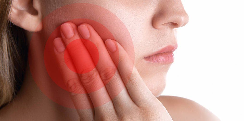
Although the etiopathophysiology of internal temporomandibular joint internal disorders (TMJ ID) is still unknown, it has been suggested that head and body posture could be related to its initial onset, development and perpetuation.

The purpose of the present study was to observe the relationship between cervical spine X-ray abnormalities and TMJ ID. This investigation evaluated 30 subjects with internal TMJ disorder symptoms (test group) and 20 healthy subjects (control group). Subjects were submitted to clinical and radiographic evaluation. Clinical evaluation comprised anamnesis and stomatognathic system physical examination. Radiographic evaluation comprised analysis of lateral cervical spine X-rays by three physical therapists and tracing on the same im ages. The test group presented twice as much cervical spine hyperlordosis as the control group (20.7% versus 10.5%), but almost half of rectification prevalence (41.4 versus 79.0%, p = 0.03). After that, the test group was divided into three subgroups according to TMJ dysfunction severity, evaluated by Helkimo's index. These subgroups were not significantly different, but the subgroup with more severe TMD showed a tendency to cervical spine hyperlordosis prevalence. Results showed a tendency for . subjects with more severe TMD to exhibit cervical spine hyperlordosis. Nevertheless, studies with a larger number of subjects suffering from severe TMD are encouraged in order to corroborate the present findings. Temporomandibular joint internal disorders (TMJ ID) are a specific category of TMJ dysfunction (TMD), a condition in which tissue morphology and healthy biodynamic joint elements are jeopardized, and which may lead to TMJ noises, altered mandibular movements, and pain. Musculoskeletal pain and altered mandibular movements may compromise the TMJ system and adjacent craniocervical region. The etiopathophysiology of TMD is still unclear. Some authors believe that head or body posture plays a role in TMD onset and development. The reasoning would be that a chronically altered craniocervical posture could lead to mandibular postural changes through biomechanical and neuromuscular mechanisms. Some authors have reported postural pattern alterations in TMD subjects in comparison with healthy subjects, while others have not found any differences between TMD and healthy subjects. SOURCE http://www.scielo.br/scielo.php?pid=s1806-83242004000400002&script=sci_arttext

La evaluación radiográfica de la columna cervical de sujetos con trastorno interno de la articulación temporomandibular

Aunque la etiopatofisiologia y de trastornos internos internos articulación temporomandibular (TMJ ID) es todavía desconocida, se ha sugerido que la cabeza y el cuerpo postura podría estar relacionada con su aparición inicial, desarrollo y perpetuación. El propósito del presente estudio fue observar la relación entre la columna cervical anomalías de rayos X y la ATM ID. Esta investigación evaluó a 30 sujetos con síntomas internos con trastorno de la ATM (grupo de prueba) y 20 sujetos sanos (grupo control). Los sujetos fueron sometidos a evaluación clínica y radiográfica. Evaluación clínica anamnesis y el examen físico que comprende el sistema estomatognático. La evaluación radiográfica compone análisis de rayos X de la columna vertebral cervical laterales por tres terapeutas físicos y el seguimiento en los mismos im edades. El grupo de prueba presenta dos veces como mucho hiperlordosis de la columna cervical como el grupo de control (20,7% frente a 10,5%), pero casi la mitad de la prevalencia de rectificación (41,4 frente a 79,0%, p = 0,03). Después de eso, el grupo de prueba se dividió en tres subgrupos en función de la gravedad de la disfunción de la ATM, evaluada por el índice de Helkimo. Estos subgrupos no fueron significativamente diferentes, pero el subgrupo con más severa TMD mostraron una tendencia a la prevalencia hiperlordosis de la columna cervical. Los resultados mostraron una tendencia a que los sujetos con más severo TMD para exhibir hiperlordosis de la columna cervical. Sin embargo, se anima a los estudios con un mayor número de sujetos que sufren de severo TMD con el fin de corroborar los resultados actuales. Trastornos de la articulación temporomandibular (ATM ID) son una categoría específica de la disfunción de la ATM (TMD), una condición en la que se ponen en peligro la morfología del tejido y elementos de unión biodinámicos saludables, y que pueden conducir a los ruidos de la articulación temporomandibular, movimientos mandibulares alteradas y dolor. El dolor musculoesquelético y los movimientos mandibulares alteradas pueden comprometer el sistema ATM y la región craneocervical adyacente. El etiopatofisiologia de TMD todavía no está claro. Algunos autores creen que la cabeza o la postura del cuerpo juega un papel en la aparición y desarrollo de TMD. El razonamiento sería que una postura craneocervical alterado crónica podría conducir a cambios posturales mandibulares mediante mecanismos biomecánicos y neuromusculares. Algunos autores han reportado alteraciones en el patrón de postura en temas de TTM en comparación con sujetos sanos, mientras que otros no han encontrado diferencias entre los TTM y sujetos sanos. SOURCE http://www.scielo.br/scielo.php?pid=s1806-83242004000400002&script=sci_arttext

By Lorenzana & Dental Associates March 6, 2019
Probablemente has escuchado en muchas ocasiones que debes tener buenos hábitos de higiene para asegurar una boca y dientes saludables. La pregunta es: ¿En qué consisten esos hábitos? Aquí te damos una explicación detallada pero simple de lo que deberías hacer: Cepillarte los Dientes:
By Lorenzana & Dental Associates January 14, 2019
¿Qué Es? El levantamiento de seno maxilar es una cirugía que agrega hueso a tu maxilar superior en el área de tus molares y premolares. El hueso es agregado entre tu maxilar y tus senos maxilares, los cuales se encuentran a cada lado de tu nariz. A modo de hacer espacio para el hueso, la membrana sinusal necesita ser levantada. ¿Para Qué Se Usa? Un levantamiento de seno se realiza cuando no hay suficiente altura en el hueso del maxilar, o cuando los senos están demasiado próximos al maxilar, y los implantes dentales no se pueden colocar por tal motivo. Hay varias posibles razones para que esto suceda: · Muchas personas que han perdido los dientes de su maxilar superior — particularmente los dientes posteriores o molares — no tienen suficiente hueso para que los implantes dentales sean colocados. Una vez se pierde un diente, el hueso en esa área comienza a ser reabsorbido. Si los dientes han estado ausentes por un largo tiempo, con frecuencia no hay suficiente hueso restante para colocar implantes. · Se pudo haber perdido hueso debido a alguna enfermedad periodontal (de las encías). · El seno maxilar puede estar demasiado próximo al maxilar superior. La forma y tamaño del seno varía de persona a persona. El seno también puede agrandarse a medida envejeces. ¿Cómo Se Hace? Tu cirujano hará una incisión y levantará la encía donde tus dientes posteriores solían estar, exponiendo el hueso. Se hace un pequeño agujero ovalado en el hueso. La membrana que reviste el seno en el lado opuesto al agujero separa tu seno de tu maxilar. Esta membrana se empuja suavemente hacia arriba y lejos de tu maxilar. Material de injerto óseo es insertado en el espacio donde el seno solía estar. Una vez el hueso está en su lugar, el tejido es suturado. Lo Que Sigue Después del procedimiento, podrías tener algo de inflamación en el área y podrías sangrar por tu boca o nariz. No te suenes la nariz o estornudes con fuerza. Hacer eso podría causar que el material de injerto óseo se mueva y que los puntos de sutura se aflojen. Puede ser que tu dentista te prescriba medicina para prevenir el congestionamiento y la inflamación. También necesitarás medicina para el dolor, un antibiótico y enjuague bucal antimicrobiano para prevenir una infección. La mayoría de los pacientes solamente experimentarán un poco de incomodidad luego de un procedimiento de levantamiento de seno. Verás al especialista luego de algunos días. Él/ella evaluará el sitio de la cirugía y retirará los puntos de sutura si fuese necesario. Podrías tener que visitar al especialista un par de veces más para que se asegure de que el área está sanando apropiadamente. Tus implantes se colocarán entre cuatro y nueve meses después del levantamiento de seno, dependiendo de la cantidad de hueso requerida. Esto permite que haya tiempo para que el material de injerto óseo se una a tu hueso.
By Lorenzana & Dental Associates January 10, 2019
Hoy en día la prótesis dental nos permite reemplazar los dientes perdidos con un aspecto muy estético y natural, devolviendo a la boca la función perdida; pero eso no fue siempre así. Las primeras prótesis dentales de las que tenemos constancia provienen de los etruscos del siglo VIII a. C. y se conserva en el Museo de la Facultad de Odontología de París. Los etruscos utilizaron dientes de animales para reemplazar las piezas faltantes ensambladas sobre bandas de oro, lo cual muestra habilidades muy altas por parte de ellos. Creemos que los fenicios ya usaron oro blando y alambre de oro para la construcción de prótesis dentales. También utilizaron soldaduras y modelos de impresiones. Hacia 754 aC , los etruscos, hábiles artesanos de la época, producían pónticos muy complejos. Empleaban bandas de oro soldadas cada una por pónticos hechos con diferentes piezas dentales de humanos o animales.
By international January 4, 2019
Aprende un poco sobre la increíble historia de la higiene dental. Learn a little about the incredible history of dental hygiene.
By international December 18, 2018
Aprende un poco sobre la increíble historia de los implantes dentales. Learn a little about the incredible history of dental implants.
By international December 12, 2018
We answer some of the most common questions about dental implants
By international December 5, 2018
CEREC is an innovative system that allows for one-visit dental restorations that are functional and esthetic. CEREC es un sistema innovador que hace posible brindar a los pacientes restauraciones dentales en un solo día, con resultados funcionales y estéticos.
By international December 1, 2018
Learn about the differences between CEREC and traditional techniques to fabricate dental restorations
By international November 29, 2018
At Lorenzana & Dental Associates, we love our CEREC system - and our patients love it, too! Thanks to CEREC, we can give people the convenience of getting their teeth fixed (e.g. crowns, veneers) in as little as a single visit to our clinic. That is just impressive, right? But how did such an incredible technology as CEREC come into existence? Well, we will talk a little about that in this blog, which is the first in a series of blogs about this system. Let's get to it!
By international November 27, 2018
Temporomandibular joint disorders are conditions that cause discomfort and inhibit the proper functioning of the jaw joint and muscles that control jaw movement. Causes In most cases, the cause of TMJ disorders is not evident. However, here is a list of potential causes: External trauma, such as a hard hit to the head or jaw. Tooth grinding or jaw clenching. Poor alignment between top and bottom teeth. Excessive chewing (e.g. gum or fingernails). Overextending the TMJ while eating large things. Degenerative diseases. Symptoms There are various symptoms that could be related to TMJ disorders. Examples of these are the following: Pain in facial, jaw, neck or back muscles. Swelling. Muscle stiffness or limited movement of the jaw. Clicking, popping or grating sounds accompanied by pain when moving the jaw. Teeth not fitting comfortably together. Jaw not opening uniformly or locking. Headache or earache. Difficulty swallowing. Diagnosis There is not always a straightforward way of diagnosing TMJ disorders. Exact causes and symptoms are not clear, and that means the discomfort someone is experimenting could be due to different health issues. Your doctor will take your symptoms and medical history into consideration and then will examine areas of discomfort, including the head, neck, face, and jaw. The doctor might also use x-rays in the analysis of your case. Treatment Reversible Treatments Conservative treatments do not invade the tissues of the face, jaw, or joint, or involve surgery. Reversible treatments do not cause permanent changes in the structure or position of the jaw or teeth. It is advisable that you try these first, before considering more expensive and permanent ones. Muscle and ligament tightness can be helped with jaw stretching and relaxing exercises as advised by your doctor. If the jaw becomes locked, it may be necessary to manipulate the joint back into place under a general anaesthetic. The most common TMJ issues are temporary and do not get worse. Short-term use of pain relievers, such as ibuprofen, may be sufficient. A stabilization splint or a bite guard may provide relief. Stabilization splints should only be used for a short time and should not cause permanent changes in bite. Permanent Treatments There is often debate around the use of permanent methods to treat TMJ disorders. There are no long term studies that show the efficacy of these methods, plus they are extensive and often more expensive than reversible treatments. The bite may be adjusted by polishing teeth, using reconstructive dentistry, or fitting braces. A repositioning splint may be fitted, which over time will permanently change the position of the jaw, ligaments and muscles. There may be an effect on teeth alignment, so more dental work may be needed to adjust the teeth. There is even the option to replace the jaw joint with artificial implants, but it should be considered a last resort. Self-Care In many cases, self-care measures can help alleviate the symptoms of TMJ disorders: Eating soft foods. Applying cold or warm compresses. Avoiding excessive jaw movements (shouting, gum-chewing, and wide yawning, for instance). Practicing gentle jaw stretching and relaxing exercises. Resting the jaw as much as possible. SOURCES: National Institute of Dental and Craniofacial Research                           Health Navigator
Show More
Share by: